Neurofunction > Volume 18(2); 2022 > Article
Jeong, Huh, and Jang: Outcomes of pallidal deep brain stimulation for treating pure blepharospasm

Abstract

Objective

Blepharospasm (BSP) is a disease in which the closure rate of the bilateral eyelids increases, mainly due to involuntary contraction of the orbicularis oculi, procerus, and corrugator muscles. The objective of this study was to report postoperative outcomes after deep brain stimulation (DBS) in 10 cases of pure BSP after at least 12 months of follow-up.

Methods

Ten patients with pure BSP who underwent bilateral globus pallidus interna (GPi) DBS at The Catholic University of Korea, Incheon St. Mary’s Hospital, between 2019 and 2021 were included. The Burke-Fahn-Marsden dystonia rating scale (BFMDRS), Blepharospasm Disability Index, and Jankovic Rating Scale were used for analysis before surgery, at 6 months of follow-up as short-term outcomes, and at follow-up over 1 year (12-37 months) as long-term results.

Results

The median age of patients at surgery was 56.5 years (interquartile range [IQR], 50.5-65.8 years) and the median length of time from disease onset to the time of surgery was 58.0 months (IQR, 46.8-64.3 months). The median postoperative follow-up period was 22.5 months (IQR, 15.3-29.0 months). The median BFMDRS movement subscale scores at the three time points (preoperative baseline, 6 months, and over 1 year of follow-up) were 7.0 (IQR, 6.0-8.0), 4.5 (IQR, 3.9-6.0; 35.7% improvement, p<0.001), and 3.8 (IQR, 2.8-5.3; 45.7% improvement, p=0.002), respectively.

Conclusion

Bilateral GPi DBS for pure BSP can be effective if conservative treatment options fail. Its benefit is not only observed in the short term, but is also maintained during long-term follow-up.

INTRODUCTION

Blepharospasm is a disease in which the closure rate of the bilateral eyelids increases, mainly due to involuntary contraction of the orbicularis oculi, procerus and corrugator muscles [1,2].
Blepharospasm, oromandibular dystonia, and Meige syndrome are different from each other but closely related. Meige syndrome is characterized by combination of cranial involvement, includes blepharospasm and involuntary oromandibular dystonia movement. Most researchers and clinicians believe that Meige syndrome, blepharospasm, and oromandibular dystonia are not one single entity, but a multi-factor origin clinical syndrome [3].
Medical treatment of these conditions has generally been unsuccessful and limited due to adverse side effects [3]. Although, botulinum toxin has been shown to be effective for many dystonic features including blepharospasm, many patients show diminished response over time [4,5]. Some patients become resistant to continuous treatment because antibodies develop [5]. To treat these disease surgically, stereotactic ablation techniques targeting the thalamus or the globus pallidus interna (GPi) have been used for several years with variable results [6-10].
The Application of deep brain stimulation (DBS) to various movement and nonmovement disorders has increased interest in the treatment of severe forms of medically refractory dystonia. In particular, the GPi was an effective target for primary generalized dystonia [11-15]. There have also been several reports of the good efficacy of GPi DBS for craniocervical dystonia or Meige syndrome [16-23].
Blepharospasm can be the most incapacitating symptom, whether focal or part of Meige syndrome. There have been rare case reports of using GPi DBS for pure blepharospasm [24,25].
The objective of this study was to report our results about postoperative outcome after DBS in total of 10 cases of pure blepharospasm after at least 12 months follow-up.

MATERIALS AND METHODS

Patients

Ten patients with pure blepharospasm who underwent GPi DBS at The Catholic University of Korea, Incheon St. Mary’s Hospital between 2019 and 2021 were included in this study. All patients were evaluated by movement disorders specialists composed of neurosurgery, neurology and rehabilitation medicine team at our center. The patients suffered severe disease-related functional impairment with unsatisfactory outcomes after oral pharmacotherapy, which included the use of benzodiazepines, anticholinergic agents, antispasmodic agents, and antiepileptic agents. All the patients also showed poor response to botulinum toxin injections. Five patients also tried some alternative therapies including physical therapy and acupuncture. Brain magnetic resonance imaging (MRI) demonstrated no structural lesion as a possible cause for their dystonia. Preoperative and postoperative Burke-Fahn-Marsden dystonia rating scale (BFMDRS) movement subscale (BFMDRS-M) scores (range 0-120), Blepharospasm Disability Index (BSDI, range 0-24), and Jankovic Rating Scale (JRS, range 0-8) were counted for each patient at 6 months and over 1 year after surgery.

Ethical statements

The present study protocol was reviewed and approved by the Institutional Review Board (IRB) of Catholic Medical Center at the Catholic University of Korea (IRB approval No. OC18RESI0108). Informed consent was obtained by all subjects when they were enrolled.

Statistical analysis

Statistical significance of changes in clinical rating scales at 6 months and over 1 year compared with baseline was assessed using the Wilcoxon signed rank test (paired). A significance threshold of 0.05 was used, and all analyses were conducted using IBM SPSS Statistics 25 (IBM Corp., Armonk, NY, USA).

Surgical procedures

All surgical procedures were performed under general anesthetics. The GPi was used for DBS target. Based on anterior and posterior commissures, initial GPi coordinates were 3 mm anterior and 3 mm inferior to the midcommissural point and 22 mm lateral to the mid-line. In addition to the above indirect targeting measures, direct targeting was implemented based on MRI performed on the day of surgery in an attempt to compensate for individual variations. We established entry/target points and the entire trajectory using planning software (Medtronic Stealthstation Planning software; Medtronic, Dublin, Ireland) to avoid the sulci, ventricle, and vessels along the trajectory. For the GPi, the trajectory was planned to be lateral to the ventricle, traverse the posterior GPi, and terminate above the optic tract. In the operative field, we usually placed the burr hole about 0.5-1 cm anterior to the coronal suture and 3.5-4 cm from the midline. Microelectrode insertion was done 3 mm above the target using three concentric bipolar tungsten microelectrodes (central, anterior, lateral) driven simultaneously by an Elekta MicroDriveTM (Elekta, Stockholm, Sweden) at incremental depths of 0.5 mm until 2 mm above the target and then 0.2 mm in depth. Intraoperative electrophysiology was performed using a LeadPoint system® (Medtronic, Tokyo, Japan) to locate and confirm GPi neuronal activity. Medtronic® 3389 quadripolar (Medtronic, Minneapolis, MN, USA) or Abbott® (Abbott Neuromodulation, Austin, TX, USA) or Boston scientific® directional electrodes (Boston Scientific, Marlborough, MA, USA) were implanted bilaterally. At a subsequent surgery, DBS electrodes were connected to an implantable pulse generator. Postoperative nonenhanced brain computed tomography imaging was performed to confirm the accuracy of electrode placement in the GPi and check postoperative complications such as intracerebral hemorrhage.

RESULTS

Clinical characteristics and individual demographic profiles of patients are summarized in Table 1. The median age of patients at surgery was 56.5 years (interquartile range [IQR], 50.5-65.8 years) and the median length of time from disease onset to the time of surgery was 58.0 months (IQR, 46.8-64.3 months). The median postoperative follow-up period was 22.5 months (IQR, 15.3-29.0 months).
Preoperatively, all the patients received medical treatments for management of their symptoms. Seven patients received benzodiazepines. One received antispasmodic drugs (baclofen). One received antiepileptic agents (topiramate). One received Gabapentin. Two received other agents such as muscle relaxant. Two received antidepressant agents (selective serotonin reuptake inhibitor, selective serotonin noradrenalin reuptake inhibitor). Eight of 10 patients (80.0%) received multiple drugs combination for managing their symptoms. All patients had an alternative treatment history with botulinum toxin injections before surgery. At the last follow-up, although all the patients were continuing medication, there was a decrease in the number or doses of medications in three of eight patients who were taking multiple drugs.
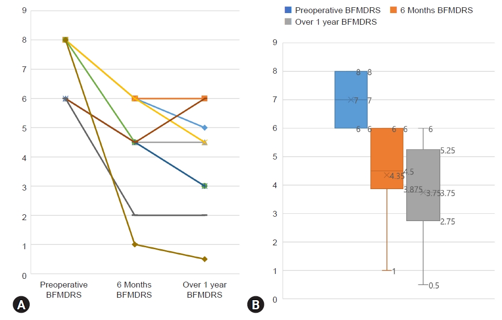
In terms of postsurgical outcome, The median of BFMDRS-M scores at the three time points (preoperative baseline, 6 months, and over 1 year of follow-up) were 7.0 (IQR, 6.0-8.0), 4.5 ([IQR, 3.9-6.0], 35.7% improvement, p<0.001), and 3.8 ([IQR, 2.8-5.3], 45.7% improvement, p=0.002), respectively (Table 2, Fig. 1). Of all patients, one showed substantial improvement in symptoms (over 80% of BFM improvement at the last follow-up). Five patients showed the best benefit at a short-term interval as well (Table 2). As compared with baseline values, the majority of movement subscores had decreased at month 6 and remained stable for over 1 year.
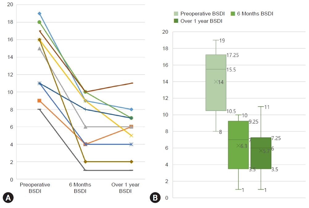
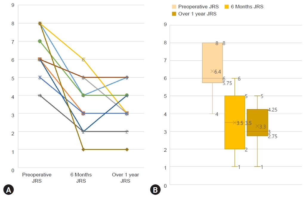
At the three time points, BSDI and JRS were checked as well. Similar to results of BFMDRS-M, improvement rate of JRS was 41.7% (p<0.001) and 50.0% (p<0.001) at 6 months and over 1 year after surgery, respectively. However, improvement rate of BSDI was 54.8% (p<0.001) and 61.3% (p<0.001) respectively for 6 months and over 1 year, slightly better than BFMDRS-M and JRS improvement rates (BSDI in Table 3, Fig. 2; JRS in Table 4, Fig. 3). All 10 patients who participated in our study had no postoperative complication such as hemorrhage and infection. There are summarized each scales and improvement rates in Table 5.
Stimulation parameters for the 10 patients are presented in Fig. 4. Of a total of 20 electrodes implanted in these 10 patients, nine patients were on a monopolar stimulation, and the rest one patient was on a bipolar stimulation at the last follow-up. The median voltage or current was 3.93 V (IQR, 3.63-4.18 V) (or mA) on the left and 3.80 V (IQR, 3.63-4.06 V) (or mA) on the right. All patients showed a stimulus intensity of more than 3 V in amplitude. The median pulse width was 75.0 μsec (IQR, 60.0-90.0 μsec) on the left and 80.0 μsec (IQR, 62.5-90.0 μsec) on the right. The median frequency was same value as 174 Hz (IQR, 162.5-180 Hz) in both sides. Six out of 10 patients were implanted using directional lead manufactured by Boston scientific® or Abbott®.

DISCUSSION

Terms of blepharospasm, oromandibular dystonia, and craniocervical dystonia are used to describe a focal or segmental dystonia whereby involuntary contraction of facial, masticatory, lingual with or without cervical muscles result in sustained and forceful movement of the related musculature. As a group of conditions that can affect the motor aspect of eye, upper and lower face, mouth and hypoglossal cranial nerves, pure blepharospasm is an uncommon disease. It is easy to make a wrong diagnosis as it can resemble symptoms of many other facial movement disorders such as hemifacial spasm, tremor, tic, chorea, and stereotypies [26]. Therefore, the diagnosis of blepharospasm can be challenging. A lot of factors including patient’s physiologic and psychological status and the clinician’s training can affect an accurate diagnosis at the time of presentation.
Since Vercueil and coworkers [27] first reported the outcome of a bilateral GPi DBS in a patient with Meige syndrome, several studies have reported that the segmental dystonia responds to GPi DBS [28-37]. However, there were only three studies on the usefulness of GPi DBS in “pure” or “isolated” blepharospasm, and even that were single case studies [24,25,38]. Our study included the largest number of patients among studies reported up to date.
According to Ostrem et al. [13], six cases with cranial-cervical dystonia showed a mean improvement rate of 86% based on the BFMDRS eyes score at 6 months postoperatively compared to baseline. Similarly, Sako et al. [33] described five patients with Meige’s syndrome who experienced 88% improvement of BFMDRS eyes score. Meanwhile, according to Reese et al. [39], 12 patients with Meige syndrome for up to 78 months after bilateral GPi DBS showed a mean eyes improvement rate of 38% after a short-term follow-up (4.4±1.5 months; p<0.001) and a rate of 47% after a long-term follow-up (38.8±21.7 months; p<0.001). Although our study was focused on pure blepharospasm, their result appeared to be similar to ours.
Santos et al. [25] reported in 2016 the first case of pallidal stimulation successfully done on pure blepharospasm, with 63% improvement rate of JRS score. This was soon followed in the same year by the report of Yamada et al. [24] of pure blepharospasm responding to bilateral pallidal DBS, with 87.5% improvement of BFMDRS score, 91.7% improvement of JRS score, 100% improvement of BSDI score, respectively. Evidente at al. [38] also reported the outcome of bilateral pallidal DBS for pure blepharospasm, with results of 100% improvement of BFMDRS and JRS score at 30 months. However, while those were all single case reports, our study is meaningful in that more patients are included.
In our study, one of patients (10.0%) had an improvement of more than 80.0% and four patients (40.0%) had 50% to 80% improvement rate compared to their baseline scores in terms of BFMDRS-M. The benefit was also sustained over the course of the last follow-up (until June 2022), although it waxed and waned depending on the patient’s condition at the day of the visit to the outpatient clinic. The results of BFMDRS-M and JRS scores were somewhat similar, but BSDI was slightly better than the others. BSDI is a quality of life form written by the patient oneself, but the other scorings are form in which the patient comes to the hospital and evaluated by the medical staff. Therefore, it is possible that the symptoms that the patient usually feels in daily routine are evaluated better than the other scores.
Moreover, the postoperative outcome could be affected to genetic or individual heterogeneity [15], and how appropriately the operation was carried out. In perspective of the operation process, the specific type of dystonia, the choice of the target, the anatomic location of the electrodes, and technical or human error during the procedure all could influence the therapeutic outcome [40-42]. Besides, various factors such as postoperative patients’ physiologic and psychologic status, inappropriate stimulation method applied during postoperative outpatient visit could limit favorable outcome. According to Wang et al. [43], Four factors such as 1) the DBS target (subthalamic nucleus vs. GPi); 2) whether symptoms first appeared at multiple sites or at a single site; 3) the subitem scores of the mouth at baseline; and 4) the follow-up period could affect significant differences between the good and poor outcome after DBS of primary Meige syndrome. Although we have yet to find predictive factors affecting the outcome, large-scale, prospective, randomized clinical trials and electrophysiological or neuroradiological studies are warranted in the future.
This study has several limitations. First, it was retrospective in nature and the outcome was collected by an unblinded rater. In addition, measures for dystonia disability scale in BFMDRS such as speech, handwriting, feeding, eating/swallowing, hygiene, dressing, and walking were not carried out. Although this should not cause a serious bias, it might have an impact on estimating the effectiveness of the treatment.

CONCLUSION

Bilateral GPi DBS in pure blepharospasm can be effective if conservative treatment options fail. The benefit is not only observed at a short-term follow-up period, but also maintained at a long-term follow-up. While no definitive risk factors affecting favorable outcome are yet to be made for pure blepharospasm, further studies in the future could clarify possible predictive factors affecting favorable outcomes. Results of this study along with those of other reports suggest that bilateral GPi DBS might be an effective treatment for medically refractory pure blepharospasm even in a long term.

NOTES

CONFLICTS OF INTEREST

No potential conflict of interest relevant to this article was reported.

Acknowledgments

We thank the staff of the Neurosurgery Department at Incheon St. Mary’s Hospital for assistance with the patient survey and video preparation.

Fig. 1.
Serial Burke-Fahn-Marsden dystonia rating scale (BFMDRS) movement subscale scores of each patient before surgery, 6 months, and over 1 year after surgery. (A) Line chart of 10 patients at baseline, 6 months, and over 1 year following deep brain stimulation (DBS). (B) Box plot of all cases at baseline, 6 months, and over 1 year following DBS.
jksfn-2022-00199f1.jpg
Fig. 2.
Serial blepharospasm Disability Index (BSDI) scores of each patient before surgery, 6 months, and over 1 year after surgery. (A) Line chart of 10 patients at baseline, 6 months, and over 1 year following deep brain stimulation (DBS). (B) Box plot of all cases at baseline, 6 months, and over 1 year following DBS.
jksfn-2022-00199f2.jpg
Fig. 3.
Serial Jankovic Rating Scale (JRS) scores of each patient before surgery, 6 months, and over 1 year after surgery. (A) Line chart of 10 patients at baseline, 6 months and over 1 year following deep brain stimulation (DBS). (B) Box plot of all cases at baseline, 6 months and over 1 year following DBS.
jksfn-2022-00199f3.jpg
Fig. 4.
Stimulation parameters and long-term BFM improvement rates for all cases. Rt: right, Lt: left, M.SC: Medtronic Activa SC, M.RC: Medtronic activa RC, B.Ge: Boston Scientific Vercise Gevia, A.In: Abbott Infinity, A.Brio: Abbott Brio, A.Libra: Abbott Libra, Amp: amplitude, PW: pulse width, BFM: Burke-Fahn-Marsden. *Directional electrode is marked with an asterisk. Each contact is indicated according to the number shown in the accompanying figure above. Stimulation with B.Ge or A.Brio/A.Libra/A.In was implemented by current-based compared to votage-based in M.SC/M.RC.
jksfn-2022-00199f4.jpg
Table 1.
Patient’s individual demographic profiles and clinical characteristics
Patient No. Sex Age at surgery (yr) Age at onset (yr) Duration of disease (mo) Medication before surgery Botulinum toxin injections before surgery Medication after surgery
1 M 50 46 59 Clonazepam, muscle relaxant + Clonazepam
2 F 50 44 76 Baclofen, clonazepam + Baclofen, clonazepam
3 F 57 53 57 SSRI, muscle relaxant + Clonazepam
4 F 52 48 59 Clonazepam + Clonazepam
5 F 70 69 23 Clonazepam, muscle relaxant + Clonazepam, alprazolam
6 M 56 53 49 Clonazepam, muscle relaxant + Clonazepam, trihexyphenidyl
7 F 68 58 127 Clonazepam, SSRI, gabapentin + Clonazepam, alprazolam
8 M 42 41 15 Clonazepam, topiramate + Clonazepam, alprazolam, procyclidine
9 F 65 60 66 Muscle relaxant + Clonazepam
10 F 66 63 46 BDZ, SSNRI + Clonazepam

M: male, F: female, SSRI: selective serotonin reuptake inhibitor, BDZ: benzodiazepine, SSNRI: selective serotonin noradrenalin reuptake inhibitor.

Table 2.
Serial BFMDRS-M scores at baseline and at 6 months and over 1 year after surgery with the overall improvement rate
Case Preoperative BFMDRS 6 months BFMDRS Over 1 year BFMDRS Improvement rate (%)
Short term Long term
1 8 6 5 25.0 37.5
2 8 6 6 25.0 25.0
3 6 4.5 4.5 25.0 25.0
4 8 6 4.5 25.0 43.8
5 6 4.5 3 25.0 50.0
6 8 4.5 3 43.8 62.5
7 6 4.5 3 25.0 50.0
8 6 4.5 6 25.0 0
9 6 2 2 66.7 66.7
10 8 1 0.5 87.5 93.8
Median (IQR) 7.0 (6.0-8.0) 4.5 (3.9-6.0) 3.8 (2.8-5.3) 35.7 (25.0-49.5) 45.7 (25.0-63.5)

BFMDRS-M: Burke-Fahn-Marsden dystonia rating scale movement subscale, IQR: interquartile range.

Table 3.
Serial BSDI scores at baseline and at 6 months and over 1 year after surgery with the overall improvement rate
Case Preoperative BSDI 6 months BSDI Over 1 year BSDI Improvement rate (%)
Short term Long term
1 19 9 8 52.6 57.9
2 9 4 6 55.6 33.3
3 15 6 6 60.0 60.0
4 16 9 5 43.8 68.8
5 11 4 4 63.6 63.6
6 18 10 7 44.4 61.1
7 11 8 7 27.3 36.4
8 17 10 11 41.2 35.3
9 8 1 1 87.5 87.5
10 16 2 2 87.5 87.5
Median (IQR) 15.5 (10.5-17.3) 7.0 (3.5-9.3) 6.0 (3.5-7.3) 54.8 (43.1-69.6) 61.3 (36.1-73.4)

BSDI: Blepharospasm Disability Index, IQR: interquartile range.

Table 4.
Serial JRS scores at baseline and at 6 months and over 1 year after surgery with the overall improvement rate
Case Preoperative JRS 6 months JRS Over 1 year JRS Improvement rate (%)
Short term Long term
1 8 4 5 50.0 37.5
2 6 3 3 50.0 50.0
3 6 5 3 16.7 50.0
4 8 6 3 25.0 62.5
5 5 3 3 40.0 40.0
6 7 4 4 42.9 42.9
7 6 2 4 66.7 33.3
8 6 5 5 16.7 16.7
9 4 2 2 50.0 50.0
10 8 1 1 87.5 87.5
Median (IQR) 6.0 (5.8-8.0) 3.5 (2.0-5.0) 3.0 (2.8-4.3) 41.7 (22.9-54.2) 50.0 (36.5-53.1)

JRS: Jankovic Rating Scale, IQR: interquartile range.

Table 5.
Median scores and improvement rates of the BFMDRS-M scores, BSDI, JRS at three time points
Scale Baseline 6 months of follow-up Over 1 year of follow-up Short-term improvement (%) Long-term improvement (%)
BFMDRS-M 7.0 (6.0-8.0) 4.5 (3.9-6.0) 3.8 (2.8-5.3) 35.7 (25.0-49.5) 45.7 (25.0-63.5)
BSDI 15.5 (10.5-17.3) 7.0 (3.5-9.3) 6.0 (3.5-7.3) 54.8 (43.1-69.6) 61.3 (36.1-73.4)
JRS 6.0 (5.8-8.0) 3.5 (2.0-5.0) 3.0 (2.8-4.3) 41.7 (22.9-54.2) 50.0 (36.5-53.1)

Values are presented as median (range).

BFMDRS-M: Burke-Fahn-Marsden dystonia rating scale movement subscale, BSDI: Blepharospasm Disability Index, JRS: Jankovic Rating Scale.

REFERENCES

1. Grandas F, Elston J, Quinn N, Marsden CD. Blepharospasm: a review of 264 patients. J Neurol Neurosurg Psychiatry 1988;51:767-72
crossref pmid pmc
2. Albanese A, Bhatia K, Bressman SB, Delong MR, Fahn S, Fung VS, et al. Phenomenology and classification of dystonia: a consensus update. Mov Disord 2013;28:863-73
crossref pmid pmc pdf
3. Ma H, Qu J, Ye L, Shu Y, Qu Q. Blepharospasm, oromandibular dystonia, and Meige syndrome: clinical and genetic update. Front Neurol 2021;12:630221
crossref pmid pmc
4. Aquino CC, Felício AC, Castro PC, Oliveira RA, Silva SM, Borges V, et al. Clinical features and treatment with botulinum toxin in blepharospasm: a 17-year experience. Arq Neuropsiquiatr 2012;70:662-6
crossref pmid
5. Colosimo C, Tiple D, Berardelli A. Efficacy and safety of long-term botulinum toxin treatment in craniocervical dystonia: a systematic review. Neurotox Res 2012;22:265-73
crossref pmid pdf
6. Andrew J, Fowler CJ, Harrison MJ. Stereotaxic thalamotomy in 55 cases of dystonia. Brain 1983;106:981-1000
crossref pmid
7. Cardoso F, Jankovic J, Grossman RG, Hamilton WJ. Outcome after stereotactic thalamotomy for dystonia and hemiballismus. Neurosurgery 1995;36:501-7. discussion 507-8
crossref pmid pdf
8. Cooper IS. 20-year followup study of the neurosurgical treatment of dystonia musculorum deformans. Adv Neurol 1976;14:423-52
pmid
9. Hassler R, Riechert T, Mundinger F, Umbach W, Ganglberger JA. Physiological observations in stereotaxic operations in extrapyramidal motor disturbances. Brain 1960;83:337-50
crossref pmid
10. Opherk C, Gruber C, Steude U, Dichgans M, Bötzel K. Successful bilateral pallidal stimulation for Meige syndrome and spasmodic torticollis. Neurology 2006;66:E14
crossref pmid
11. Kumar R, Dagher A, Hutchison WD, Lang AE, Lozano AM. Globus pallidus deep brain stimulation for generalized dystonia: clinical and PET investigation. Neurology 1999;53:871-4
crossref pmid
12. Kupsch A, Benecke R, Müller J, Trottenberg T, Schneider GH, Poewe W, et al; Deep-Brain Stimulation for Dystonia Study Group. Pallidal deep-brain stimulation in primary generalized or segmental dystonia. N Engl J Med 2006;355:1978-90
crossref pmid
13. Ostrem JL, Marks WJ Jr, Volz MM, Heath SL, Starr PA. Pallidal deep brain stimulation in patients with cranial-cervical dystonia (Meige syndrome). Mov Disord 2007;22:1885-91
crossref pmid
14. Tronnier VM, Fogel W. Pallidal stimulation for generalized dystonia. Report of three cases. J Neurosurg 2000;92:453-6
crossref pmid
15. Vidailhet M, Vercueil L, Houeto JL, Krystkowiak P, Benabid AL, Cornu P, et al; French Stimulation du Pallidum Interne dans la Dystonie (SPIDY) Study Group. Bilateral deep-brain stimulation of the globus pallidus in primary generalized dystonia. N Engl J Med 2005;352:459-67
crossref pmid
16. Bereznai B, Steude U, Seelos K, Bötzel K. Chronic high-frequency globus pallidus internus stimulation in different types of dystonia: a clinical, video, and MRI report of six patients presenting with segmental, cervical, and generalized dystonia. Mov Disord 2002;17:138-44
crossref pmid pdf
17. Blomstedt P, Tisch S, Hariz MI. Pallidal deep brain stimulation in the treatment of Meige syndrome. Acta Neurol Scand 2008;118:198-202
crossref pmid
18. Capelle HH, Weigel R, Krauss JK. Bilateral pallidal stimulation for blepharospasm-oromandibular dystonia (Meige syndrome). Neurology 2003;60:2017-8
crossref pmid
19. Foote KD, Sanchez JC, Okun MS. Staged deep brain stimulation for refractory craniofacial dystonia with blepharospasm: case report and physiology. Neurosurgery 2005;56:E415; discussion E415
crossref pmid pdf
20. Hebb MO, Chiasson P, Lang AE, Brownstone RM, Mendez I. Sustained relief of dystonia following cessation of deep brain stimulation. Mov Disord 2007;22:1958-62
crossref pmid
21. Loher TJ, Capelle HH, Kaelin-Lang A, Weber S, Weigel R, Burgunder JM, et al. Deep brain stimulation for dystonia: outcome at long-term follow-up. J Neurol 2008;255:881-4
crossref pmid pdf
22. Markaki E, Kefalopoulou Z, Georgiopoulos M, Paschali A, Constantoyannis C. Meige’s syndrome: a cranial dystonia treated with bilateral pallidal deep brain stimulation. Clin Neurol Neurosurg 2010;112:344-6
crossref pmid
23. Muta D, Goto S, Nishikawa S, Hamasaki T, Ushio Y, Inoue N, et al. Bilateral pallidal stimulation for idiopathic segmental axial dystonia advanced from Meige syndrome refractory to bilateral thalamotomy. Mov Disord 2001;16:774-7
crossref pmid
24. Yamada K, Shinojima N, Hamasaki T, Kuratsu J. Pallidal stimulation for medically intractable blepharospasm. BMJ Case Rep 2016;2016:bcr2015214241
crossref pmid pmc
25. Santos AF, Veiga A, Augusto L, Vaz R, Rosas MJ, Volkmann J. Successful treatment of blepharospasm by pallidal neurostimulation. Mov Disord Clin Pract 2016;3:409-11
crossref pmid pmc pdf
26. Evidente VG, Adler CH. Hemifacial spasm and other craniofacial movement disorders. Mayo Clin Proc 1998;73:67-71
crossref pmid
27. Vercueil L, Pollak P, Fraix V, Caputo E, Moro E, Benazzouz A, et al. Deep brain stimulation in the treatment of severe dystonia. J Neurol 2001;248:695-700
crossref pmid pdf
28. Lyons MK, Birch BD, Hillman RA, Boucher OK, Evidente VG. Long-term follow-up of deep brain stimulation for Meige syndrome. Neurosurg Focus 2010;29:E5
crossref
29. Sensi M, Cavallo MA, Quatrale R, Sarubbo S, Biguzzi S, Lettieri C, et al. Pallidal stimulation for segmental dystonia: long term follow up of 11 consecutive patients. Mov Disord 2009;24:1829-35
crossref pmid pdf
30. Woehrle JC, Blahak C, Kekelia K, Capelle HH, Baezner H, Grips E, et al. Chronic deep brain stimulation for segmental dystonia. Stereotact Funct Neurosurg 2009;87:379-84
crossref pmid pdf
31. Inoue N, Nagahiro S, Kaji R, Goto S. Long-term suppression of Meige syndrome after pallidal stimulation: a 10-year follow-up study. Mov Disord 2010;25:1756-8
crossref pmid
32. Romito LM, Elia AE, Franzini A, Bugiani O, Albanese A. Low-voltage bilateral pallidal stimulation for severe meige syndrome in a patient with primary segmental dystonia: case report. Neurosurgery 2010;67:onsE308; discussion onsE308
crossref pmid
33. Sako W, Morigaki R, Mizobuchi Y, Tsuzuki T, Ima H, Ushio Y, et al. Bilateral pallidal deep brain stimulation in primary Meige syndrome. Parkinsonism Relat Disord 2011;17:123-5
crossref pmid
34. Tai CH, Wu RM, Liu HM, Tsai CW, Tseng SH. Meige syndrome relieved by bilateral pallidal stimulation with cycling mode: case report. Neurosurgery 2011;69:E1333-7
crossref pmid
35. Sobstyl M, Ząbek M, Mossakowski Z, Zaczyński A. Pallidal deep brain stimulation in the treatment of Meige syndrome. Neurol Neurochir Pol 2014;48:196-9
crossref pmid
36. Wang X, Zhang C, Wang Y, Liu C, Zhao B, Zhang JG, et al. Deep brain stimulation for craniocervical dystonia (Meige syndrome): a report of four patients and a literature-based analysis of its treatment effects. Neuromodulation 2016;19:818-23
crossref pmid pdf
37. Berman BD, Starr PA, Marks WJ Jr, Ostrem JL. Induction of bradykinesia with pallidal deep brain stimulation in patients with cranial-cervical dystonia. Stereotact Funct Neurosurg 2009;87:37-44
crossref pmid pmc pdf
38. Evidente VGH, Ponce FA, Evidente MH, Lambert M, Garrett R. Tardive blepharospasm may respond to bilateral pallidal deep brain stimulation. Tremor Other Hyperkinet Mov (N Y) 2021;11:10
crossref pmid pmc
39. Reese R, Gruber D, Schoenecker T, Bäzner H, Blahak C, Capelle HH, et al. Long-term clinical outcome in meige syndrome treated with internal pallidum deep brain stimulation. Mov Disord 2011;26:691-8
crossref pmid pdf
40. Cif L, El Fertit H, Vayssiere N, Hemm S, Hardouin E, Gannau A, et al. Treatment of dystonic syndromes by chronic electrical stimulation of the internal globus pallidus. J Neurosurg Sci 2003;47:52-5
pmid pdf
41. Volkmann J, Benecke R. Deep brain stimulation for dystonia: patient selection and evaluation. Mov Disord 2002;17 Suppl 3:S112-5
crossref pmid
42. Li Z, Zhang JG, Ye Y, Li X. Review on factors affecting targeting accuracy of deep brain stimulation electrode implantation between 2001 and 2015. Stereotact Funct Neurosurg 2016;94:351-62
crossref pmid pdf
43. Wang X, Mao Z, Cui Z, Xu X, Pan L, Liang S, et al. Predictive factors for long-term clinical outcomes of deep brain stimulation in the treatment of primary Meige syndrome. J Neurosurg 2019;132:1367-75
crossref pmid


ABOUT
BROWSE ARTICLES
EDITORIAL POLICY
FOR CONTRIBUTORS
Editorial Office
Department of Neurosurgery, Yonsei University College of Medicine
50 Yonsei-ro, Seodaemun-gu, Seoul 03722, Korea
Tel: +82-2-2228-2150    Fax: +82-2-393-9979    E-mail: changws@yonsei.ac.kr / changws0716@yuhs.ac                

Copyright © 2024 The Korean Society of Stereotactic and Functional Neurosurgery.

Developed in M2PI

Close layer
prev next